11 Focused Assessment- Respiratory System
Learning Objectives
At the end of this chapter, the learner will:
- Conduct a health history pertaining to the respiratory system.
- Identify anatomic landmarks in identifying underlying structures and the location of physical findings.
- Inspect the thorax for pattern of respiration, skin, symmetry, and use of accessory muscles.
- Auscultate the anterior and posterior thorax for normal breath sounds and adventitious sounds.
- Describe the findings using correct terminology.
- Document the findings of the respiratory exam.
I. Overview of the Respiratory System
The assessment of the respiratory system includes assessing the thorax, lungs, ventilatory function, and oxygenation of the body. Focused assessment techniques are applied intensively in this process, including: inspecting the level of consciousness, agitation, skin color, clubbing of fingers, shortness of breath, use of accessory muscles, and position and alignment of the spine; auscultating breathing sounds; palpating the position of the trachea and checking for subcutaneous emphysema; percussing the chest to assess the underlying structure.
II. Anatomy and Physiology
Identifying thoracic landmarks is essential to the systematic examination of the respiratory system. Click the link below to review anatomy and physiology of the respiratory system. You will need to apply your knowledge in the assessment process.
III. Medical Terminology
Important terms to know and understand:
| Adventitious sounds | abnormal breath sounds |
| Apnea | involuntary cessation of breathing |
| Atelectasis | incomplete expansion or collapse of a part of the lungs |
| Bradypnea | slow rate of breathing |
| Cheyne-Stokes Respirations | gradual increase and then gradual decrease in depth of respiration followed a period of apnea |
| Crackles | crackling sounds made as air moves through wet secretions in the lungs |
| Crepitus | a grating sound or sensation under the skin around the lungs, or in the joints |
| Cyanosis | bluish coloring of the skin |
| Dyspnea | difficult or labored breathing |
| Hemoptysis | sputum containing blood |
| Hyperventilation | condition in which there is more than the normal amount of air entering and leaving lungs |
| Hypoventilation | decreased rate or depth of air movement into the lungs |
| Hypoxia | inadequate amount of oxygen available to the cells |
| Nasal Flaring | nostrils widen while breathing indicates difficulty in breathing |
| Orthopnea | shortness of breath when lying flat and relieved by sitting or standing |
| Pneumothorax | air in the pleural space |
| Stridor | harsh, high-pitched sound usually heard on inspiration when upper airway become narrowed |
| Tachypnea | rapid rate of breathing |
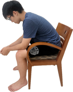
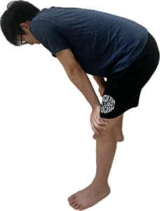
| Tripod Position or Orthopneic Position | a person sits or stands leaning forward and supports the upper body with hands on knees or other surface, often adopted by people experiencing respiratory distress |
| Wheezes | high-pitched, musical noise that sounds like a squeak |
IV. Step by Step Assessment
Safety considerations:
- Perform hand hygiene.
- Check room for contact precautions.
- Introduce yourself to patient.
- Confirm patient ID using two patient identifiers (e.g., name and date of birth).
- Explain process to patient.
- Be organized and systematic in your assessment.
- Use appropriate listening and questioning skills.
- Listen and attend to patient cues.
- Ensure patient’s privacy and dignity.
- Apply principles of asepsis and safety.
- Check vital signs.
| Steps | Additional Information |
| 1. Conduct a focused interview related to the history of respiratory disease, smoking, and environmental exposures. | Ask relevant questions related to dyspnea, cough/sputum, fever, chills, chest pain with breathing, and previous history such as allergies, asthma, chronic obstructive pulmonary disease (COPD), or pneumonia. Inquire about home respiratory equipment such as oxygen, nebulizer devices, CPAP, or BiPAP, as well as related medications or supplements.
Note: CPAP and BiPAP are devices used to treat sleep-related breathing problems like sleep apnea, which cause cessation or pauses in breathing while asleep. These devices deliver airflow to maintain an open airway. |
2. Inspect:
|
 Asymmetrical chest expansion may indicate conditions such as pneumothorax, rib fracture, severe pneumonia, or atelectasis. With hypoxemia, cyanosis of the extremities or around the mouth may be noted. |
3. Auscultate lungs for breath sounds and adventitious sounds.
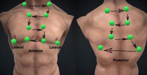
|
Fine crackles (rales) may indicate asthma and chronic obstructive pulmonary disease (COPD). Example below.
Coarse crackles may indicate pulmonary edema. Wheezing may indicate asthma, bronchitis, or emphysema. Example below. Low-pitched wheezing (rhonchi) may indicate pneumonia. Pleural friction rub (creaking) may indicate pleurisy or pleuritis. Stridor is a high-pitched breath sound caused by a narrowed or obstructed airway. Example below. Interventions should be provided if a decrease or absence of breath sounds is noted, as it may indicate severe respiratory problems such as airway obstruction, pneumonia, pneumothorax, pleural effusion, or atelectasis (Reyes et al., 2020). |
| 4. Report and document assessment findings and related health problems according to agency policy. | Accurate and timely documentation and reporting promote patient safety. |
A more detailed overview of respiratory examination is available at: Lung Exam (text) and Respiratory Sounds (text and video)
Knowledge Check
V. Documentation of Assessment Findings
A sample narrative documentation:
A & O x 4, denies shortness of breath or chest pain, RR18, without use of accessory muscles, symmetrical chest wall movement, clear breath sounds in all lung fields, O2 98% in room air.
VI. Related Laboratory and Diagnostic Procedures/ Findings
- Spirometry Test (Pulmonary Function Test): This test involves having the patient inhale and exhale through a device to measure lung capacity. It is used to diagnose conditions such as asthma and chronic obstructive pulmonary disease (COPD).
- Chest X-ray: This imaging test provides a view of the structures inside the chest and is a useful test for diagnosing pneumonia and other lung abnormalities.
- Computerized Tomography (CT) Scan: A CT scan offers detailed cross-sectional images of the chest and can identify respiratory issues that may not be visible on a standard X-ray.
- Bronchoscopy: This invasive procedure involves inserting a fiberscope into the airway to examine the bronchi. It is used to retrieve tissue samples (biopsy) for diagnosing lung cancer and to treat airway blockages or obstructions caused by foreign objects.
VII. Learning Exercises
VIII. Attributions and References
- A.D.A.M. Medical Encyclopedia [Internet]. Johns Creek (GA): Ebix, Inc., A.D.A.M.; c1997-2020. Nail abnormalities; [updated 2019 Jul 31; reviewed 2019 Apr 16; cited 2020 Aug 30]; [about 4 p.]. Available from: https://medlineplus.gov/ency/article/003247.htm
- Anderson, R., Doyle, G. R., & McCutcheon, J. A. Clinical procedures for safer patient care. https://pressbooks.bccampus.ca/clinicalproceduresforsaferpatientcaretrubscn/chapter/2-7-head-to-toe-assessment-chest-respiratory-assessment/ Accessed July 4th, 2021.
- Deviant Art: Male torso render image by Illtrytobeapro. Oct. 7, 2013. ODI: https://www.deviantart.com/illtrytobeapro/art/Male-Torso-render-405887318 Accessed July 4th, 2021.
- Doyle, G. R. & McCutcheon, J. A. Step by Step Checklist adapted from https://opentextbc.ca/clinicalskills/chapter/2-5-focussed-respiratory-assessment/
- Khan Academy: Meet the lungs by Rishi Desai. https://www.khanacademy.org/science/high-school-biology/hs-human-body-systems/hs-the-circulatory-and-respiratory-systems/v/meet-the-lungs
- Pinto VL, Sharma S. Continuous Positive Airway Pressure. [Updated 2023 Jul 24]. In: StatPearls [Internet]. Treasure Island (FL): StatPearls Publishing; 2024 Jan-. Available from: https://www.ncbi.nlm.nih.gov/books/NBK482178/
- Reyes, F.M. Modi, P., & Le, J.K. (Updated July 10, 2020). Lung Exam. In: StatPearls [Internet]. Treasure Island (FL): StatPearls Publishing; 2021 Jan. DOI: https://www.ncbi.nlm.nih.gov/books/NBK459253/ Accessed July 5th, 2021.
- Wikipedia contributors. (2019, July 30). Respiratory sounds. In Wikipedia, The Free Encyclopedia. Retrieved 23:46, August 29, 2019, from https://en.wikipedia.org/w/index.php?title=Respiratory_sounds&oldid=908541418
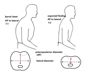
- Wikimedia Commons. Barrel chest adapted from DOI: https://commons.wikimedia.org/wiki/File:%CE%92%CF%85%CF%84%CE%B9%CE%BF%CE%B5%CE%B9%CE%B4%CE%AE%CF%82_%CE%B8%CF%8E%CF%81%CE%B1%CE%BA%CE%B1%CF%82_(barrel_chest).png
