12 Focused Assessment- Gastrointestinal and Genitourinary
Learning Objectives
At the end of this chapter, the learner will:
- Provide safety and privacy during gastrointestinal and genitourinary assessments.
- Identify the location of the organs contained in the abdominal cavity.
- Obtain health history relevant to the gastrointestinal and genitourinary assessments.
- Describe two methods (4 quadrants and 9 regions) of anatomic mapping used to describe findings related to the abdominal assessment.
- Perform gastrointestinal and genitourinary assessments using the correct exam order and techniques.
- Document findings of the gastrointestinal and genitourinary systems using correct medical terminology.
I. Overview of the gastrointestinal (GI) and Genitourinary (GU) Systems:
The gastrointestinal (GI) system is responsible for the ingestion of food, the absorption of nutrients, and eliminating waste as stool. The genitourinary (GU) system, also called the renal or urinary system is responsible for electrolytes and pH balance, blood volume and pressure, and eliminates waste as urine.
II. Anatomy and Physiology
Please use the following videos and other learning tools to review the Anatomy and Physiology of the Gastrointestinal system and Genitourinary systems. The first picture is of all organs within the gastrointestinal system. Use this picture to identify organs within each abdominal quadrant or within each region when utilizing the four quadrants and/or the 9 region methods to systematically exam the abdomen and/or document the findings.
Gastrointestinal System


The video below demonstrates peristalsis in the large intestines.
The GI tract has three priority functions: digestion of food, absorption of nutrients, and elimination of waste. Digestion involves breaking down into its smallest parts so nutrients can be absorbed. There are two types of digestion. The first is mechanical and this process is completed in the mouth and stomach by chewing food into smaller pieces. Chemical digestion occurs later and chemicals are used to break down food molecules even more to even smaller molecules for easier absorption. Absorption occurs when nutrients pass through to the bloodstream or lymph system and is circulated throughout the body. Anything not digested and absorbed is moved to the anus for elimination.
The small intestine is the area in which most digestion occurs. There is the completion of proteins and carbohydrates digestion and lipids are solely digested in the small intestine. Most of the water from food is reabsorbed here. There are three sections called the duodenum, jejunum, and Ileum.
Please click on this link for further information about the small intestine: https://openstax.org/books/anatomy-and-physiology/pages/23-5-the-small-and-large-intestines
The large intestine also called the Colon processes chyme, reabsorbs water, electrolytes, and vitamins from waste prior to it becoming feces. It also produces vitamins B and K . There are four parts to the large intestine and these include the cecum, the ascending colon, transverse colon, descending colon, and the sigmoid colon.
Please click on this link for further information about the large intestine: https://www.ncbi.nlm.nih.gov/books/NBK507857/
The Genitourinary System:
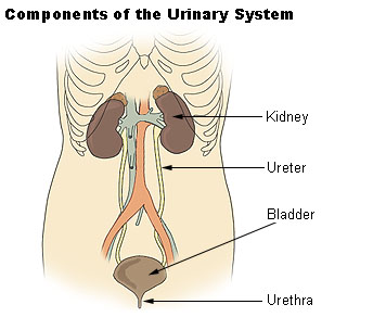
The GU system is made up of the kidneys, ureters, urinary bladder, and urethra. This system is responsible for:
- regulating pH
- synthesizing vitamin D
- producing erythropoietin
- cleaning the blood of waste
- reabsorbing sodium, water, glucose, amino acids, and vitamins
- produces renin for the Aldosterone-Renin-Angiotensin system
- producing urine to rid the body of waste. The GU system includes the kidneys, ureters, urinary bladder, and urethra.
Please click on this link for further information about the GU system: https://courses.lumenlearning.com/nemcc-ap/chapter/gross-anatomy-of-the-kidney/
Knowledge Check
III. Medical Terminology
| Ascites | accumulation of fluid in the peritoneal cavity |
| Anuria | no urine; 24-hour urine output is less than 100 mL |
| Azotemia | high levels of nitrogen-containing compounds (body waste) in the blood |
| Blood urea nitrogen (BUN) | a blood test to check the amount of urea nitrogen |
| Creatinine | a waste generated from muscle metabolism |
| Diuresis | increase in urine excretion |
| Frequency | increase number of urinations |
| Gastroesophageal Reflux Disease (GERD) | Stomach acid flows up from the stomach into the esophagus causing heartburn |
| Hematemesis | vomiting blood |
| Hesitancy | Inability to start or maintain a urine stream |
| Hematuria | Blood in urine |
| Hiatal Hernia | the protrusion of the stomach through the esophageal opening in the diaphragm |
| Hypervolemia | too much fluid in the blood |
| Hypovolemia | a decreased volume of circulating blood in the body. |
| Incontinence | loss of bladder control leading to involuntary urination |
| Melena | dark tarry sticky feces containing blood |
| Oliguria | inadequate production or secretion of urine; 24-hour urine output less than 400 mL |
| Peristalsis | the involuntary constriction and relaxation of the muscles of the intestine |
| Peritoneum | the serous membrane lining the cavity of the abdomen and covering the abdominal organs |
| Polyuria | the excessive output of urine (diuresis) |
| Rebound Tenderness | pain felt when pressure as to the abdomen is suddenly removed |
| Urea | a nitrogen-containing substance normally cleared from the blood by the kidney into the urine |
| Urinary Retention | inability to empty the bladder completely |
| Urinary Stasis | Urine flow has been impaired and is used interchangeably with urinary retention. |
| Urinary Tract Infection (UTI) | an infection in the urinary system |
Knowledge Check
Assessment of the gastrointestinal system includes obtaining relevant subjective and objective data and is focused on the mouth and abdomen. The genitourinary system assessment also needs subjective and objective data and focuses on the examination of the body’s ability to get rid of urine, the genitalia, and the urine itself.
Pain is the most common complaint related to abdominal problems and can be attributed to multiple underlying etiologies. Because of the potential variability of contributing factors, a careful and thorough assessment of this chief complaint should occur.
Nausea, vomiting, diarrhea, and constipation are common issues experienced by hospitalized patients due to adverse effects of medications or medical procedures. It is important to ask a hospitalized patient daily about the date of their last bowel movement and flatus so that a bowel management program can be initiated if necessary. If a patient is experiencing diarrhea, it is important to assess and monitor for signs of dehydration or electrolyte imbalances. Dehydration can be indicated by dry skin, dry mucous membranes, or sunken eyes. These symptoms may require contacting the health care provider for further treatment.
Additional specialized assessments of GI system function can include examination of the oropharynx and esophagus. For example, patients who have experienced a cerebrovascular accident (CVA), also called a “stroke,” may experience difficulty swallowing (dysphagia). The nurse is often the first to notice these difficulties when swallowing pills, liquid, or food and can advocate for treatment to prevent complications, such as unintended weight loss or aspiration pneumonia.[4]
The nursing assessment of the genitourinary system generally focuses on bladder function. Ask about urinary symptoms, including, urinary frequency, or urinary urgency. Dysuria is any discomfort associated with urination and often signifies a urinary tract infection. Patients with dysuria commonly experience burning, stinging, or itching sensations. In elderly patients, changes in mental status may be the presenting symptom of a urinary tract infection. In women with dysuria, asking whether the discomfort is internal or external is important because vaginal inflammation can also cause dysuria as the urine passes by the inflamed labia.
Abnormally frequent urination (e.g., every hour or two) is termed urinary frequency. In older adults, urinary frequency often occurs at night and is termed nocturia. The frequency of normal urination varies considerably from individual to individual depending on personality traits, bladder capacity, or drinking habits. It can also be a symptom of a urinary tract infection, pregnancy in females, or prostate enlargement in males.
Urinary urgency is an abrupt, strong, and often overwhelming need to urinate. Urgency often causes, a leakage of urine. When patients experience urinary urgency, the desire to urinate may be constant with only a few milliliters of urine eliminated with each voiding.[5
IV. Step by Step Assessment
- Perform hand hygiene.
- Check room for contact precautions.
- Introduce yourself to the patient.
- Confirm patient ID using two patient identifiers (e.g., name and date of birth).
- Explain the process to the patient.
- Assemble equipment prior to starting the exam.
- Be organized and systematic in your assessment.
- Use appropriate listening and questioning skills.
- Listen and attend to patient cues.
- Ensure patient’s privacy and dignity.
- Apply principles of asepsis and safety.
- Check vital signs.
| Steps |
Additional Information |
| 1. Conduct a focused interview related to the Gastrointestinal system. | Ask relevant questions to obtain subjective data relevant to:
|
2. Inspect:
|
|
3. Auscultate the abdomen in all four quadrants
|
|
4. Palpate the abdomen
|
In the MC nursing program, percussion is considered an advanced practice skill and is not an expectation for students. Therefore, the next step of the abdominal assessment will be palpation.
Nurses complete light palpation only. All four quadrants need to be palpated.
|
| 5. Report and document assessment findings and related health problems according to agency policy. | Accurate and timely documentation and reporting promote patient safety. |


| Steps |
Additional Information |
| 1. Conduct a focused interview related to the Genitourinary system. | Ask relevant questions to obtain subjective data relevant to:
|
Inspect
|
|

A sample of narrative documentation
Sample of normal findings:
The patient denies any abdominal pain or change to bowel habits has a soft formed brown stool once a day. Denies any loss of appetite or change in weight. Denies any difficulty chewing or swallowing or any family history of abdominal cancers. The abdomen is the same color as the rest of the body with sparse hair distribution close to the perineum. The abdomen is rounded, symmetrical with straie around the waist. The umbilicus is midline and no odor or discharge noted. The abdomen is soft, non-distended, non-tender with positive bowel sounds to all four quadrants and no guarding noted during the assessment.
The patient denies any change to the urinary pattern and urinates 4-8 times a day, denies frequency, hesitancy, dysuria, hematuria, urgency, or incontinence. Denies any flank pain or change n appetite or energy. Abdomen flat and symmetrical, midline umbilicus, no scars, skin color consistent with the rest of the body. No distention to the suprapubic area and no edema to face, hand, or lower extremities noted.
VI. Related Laboratory and Diagnostic Procedures/Findings
Abdominal pain is the most common chief complaint related to abdominal assessment. This pain can be associated with the following: stomach/ intestine (digestion) problems, liver/gall bladder issues, pancreas problems, appendicitis, urinary tract problems, and or problems from the reproductive system. Therefore, in addition to information gathered from health history and physical assessment, some laboratory tests and diagnostic exams can assist in diagnosing the cause of the pain.
A complete blood count (CBC), is a blood test that can help determine if an infection is present in the body, like UTI (urinary tract infection) or peritonitis (infection of peritoneum).
Urinalysis is a urine test to determine if blood or infection is present in the urinary tract and may be ordered if the patient presents with pain to the abdomen, pelvis, back, or flank areas to check for a Urinary Tract Infection (UTI)
Liver Function Test (LFT), is a blood test to check the levels of liver enzymes. When liver enzymes are elevated the liver has been damaged and liver function may beimpaired.
Amylase and Lipase are blood tests to check the enzyme levels produced by the pancreas. Patients with pancreatitis will have elevated levels of these two enzymes.
Occult Stool/Hemoccult Test is a stool test. It checks the blood in stool that cannot be seen with the bare eye. Positive results indicate a problem (may be bleeding) in the gastrointestinal (GI) tract.
An X-ray of the abdomen (KUB) examines the kidneys, ureters, bladder, intestines, and the bones of the pelvis and spine. The following are seen on this type of XRAY: kidney stone, gas in the GI tract, or constipation.
Ultrasound uses sound waves to create images. It is often used to diagnose problems with the gallbladder or the kidneys
Computed tomography (CT) scan is used to create more detailed images of the body.
Magnetic resonance imaging (MRI) uses magnetic fields to produce images of the body. However, it cannot be performed in patients who have metal in their bodies.
Esophagogastroduodenoscopy (EGD) uses an endoscope with a camera is inserted and progressed through the patient’s mouth, to the esophagus, to the stomach, and finally to the duodenum. This test is used to directly inspect the GI tract (up to the duodenum). This test will help diagnose acid reflux, ulcers, or mass (cancer).
Colonoscopy, like EGD, is a test to examine the inside of the colon with an endoscope. This test will help to diagnose cancer, diverticulitis (infection of small, bulging pouches in the digestive tract), and colitis.
A more detailed overview of about the GI and GU assessment please click on this link: https://wtcs.pressbooks.pub/nursingskills/chapter/12-3-gastrointestinal-and-genitourinary-assessment/
VII. Learning Exercises
VIII. Attributions and References
Narayan, R. https://www.khanacademy.org/science/high-school-biology/hs-human-body-systems/hs-the-digestive-and-excretory-systems/v/meet-the-gastrointestinal-tract
Narayan, R. https://www.khanacademy.org/science/high-school-biology/hs-human-body-systems/hs-the-digestive-and-excretory-systems/v/how-do-our-kidneys-work
Human Physiology/The Urinary System. Provided by: Wikibooks. Located at: http://en.wikibooks.org/wiki/Human_Physiology/The_Urinary_System%23Introduction. License: CC BY-SA: Attribution-ShareAlike
Nursing Skills by Open Resources for Nursing (Open RN) 12.3 Gastrointestinal and Genitourinary Assessment by Wisconsin Technical College System Retrieved at 18:19 July 23, 2021 wtcs.pressbooks.pub/nursingskills/chapter/12-3-gastrintestinal-and-genitourinary-assessment/#footnote-385-1 is licensed under a Creative Commons Attribution 4.0 International License, except where otherwise noted
Overview of the Urinary System by Lumen Boundless Anatomy and Physiology Retrieved 18:30 at July 23, 2021, https://courses.lumenlearning.com/boundless-ap/chapter/overview-of-the-urinary-system/Provided by: Boundless.com. License: CC BY-SA: Attribution-ShareAlike
Introduction to the Digestive System by biology LibreTexts retrieved ar 20:00 July 23,2021 3 D Medical Animation-Peristalsis in Large Intestine https://youtu.be/Ujr0UAbyPS4
File: There are (a) nine abdominal regions and (b) four abdominal quadrants in the peritoneal cavity (2013, June 3). Wikimedia Commons, the free media repository. Retrieved 20:12, July 23, 2021, from https://commons.wikimedia.org/wiki/File:Abdominal_Quadrant_Regions.jpg
Anatomy & Physiology by Oregon State University 23.1 Overview of the digestive SystemAnotomy & Physiology provided by open Oregon state education retrieved at 21:30 July 23,2021 https://open.oregonstate.education/aandp/chapter/23-1-overview-of-the-digestive-system/
Step by Step Checklist adapted from https://opentextbc.ca/clinicalskills/chapter/2-5-focussed-respiratory-assessment/ by authors Glynda Rees Doyle and Jodie Anita McCutcheon
WikiDoc editors: Gibson, C. M. & Zorkun, C. (2015). Decreased bowel sounds. Retrieved 17:36, March 25, 2021. from https://www.wikidoc.org/index.php/Decreased_bowel_sounds#cite_ref-1
Wikipedia contributors. (2019, April 8). Abdominal examination. In Wikipedia, The Free Encyclopedia. Retrieved 01:19, August 30, 2019, from https://en.wikipedia.org/w/index.php?title=Abdominal_examination&oldid=891511595
Wisconsin Technical College System 12.3 Gastrointestinal and Genitourinary Assessment one resources for Nursing (open RN) Retrieved 0845 July 29 2021 https://wtcs.pressbooks.pub/nursingskills/chapter/12-3-gastrointestinal-and-genitourinary-assessment/
